Abstract
Non-invasive measurements of scalp electric potentials (EEG) and of magnetic fields (MEG) are generated by the primary current density distribution1 that arises from neuronal post-synaptic processes. A solution to the inverse problem2 (i.e., current density estimation) is of general interest, since high time resolution images of electric neuronal activity would provide significant information on the time course and the localization of brain function. However, this problem has no unique solution, and all previous linear-distributed solutions3-5 at best produced low spatial resolution images with systematic localization errors. We present a new linear-distributed solution that yields images of statistically standardized current density with exact point-wise localization.
Results
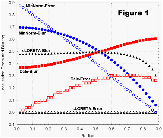
In a theoretical setting, a unit-radius conducting sphere was considered, with exact known electric potential distribution over the entire surface, and with totally unknown current density vector field within the volume with radius smaller than 0.9. Exact equations were derived for sLORETA, the minimum norm3 (MN), and the Dale et al.5 methods. Figure 1 shows localization errors and spatial dispersions (blurring) as a function of the radius of the test point source. The results were similar for a very wide range of the regularization parameter (which corrects for possible noise in the measurements). Log10-ratios of estimated source strength (shallow to deep) were 4.9, 1.0, and, 2.4, for MN, Dale, and sLORETA, respectively. sLORETA always localized exactly.
In a more practical setting, by means of numerical computations, the method was tested in a realistic head model, in one case with unknown current density (unknown orientation) for 6430 voxels distributed throughout a blurred representation of cortical grey matter volume, and in the second case with unknown current density (known orientation) for 12980 triangles distributed over the cortical surface6. EEG and MEG were tested separately, with as few as 25 electrodes/sensors, with and without noise in measurements. In the no-noise condition, sLORETA always had exact localization (zero error) for test point sources. In the noise condition, with appropriate regularization, sLORETA yielded the lowest localization errors of all three tomographies.
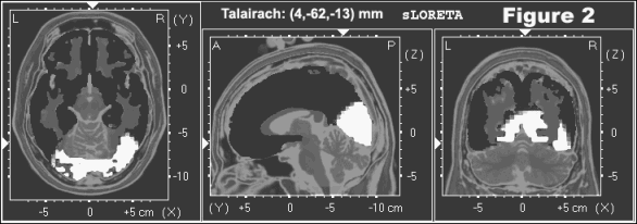
Finally, a simple empirical validation test was carried out on sLORETA, by analyzing event related responses to visual stimulation with pictures of human faces, using 25 electrodes, in 17 subjects. Figure 2 shows a statistical non-parametric map at latency 167ms, demonstrating significant activation in Brodmann areas 18/17 and right 37 (fusiform gyrus).
Conclusion
Under a diverse range of implementations, the new tomography sLORETA is exceptionally capable of exact (zero error) localization when evaluated with point sources. Barring low spatial resolution, which worsens with depth, the method is potentially capable of producing low resolution images for any current density that can be represented as a numerable set of "hot spots".
References
1. Hämäläinen, M.S. et al. Rev. Mod. Phys. 65, 413-497 (1993).
2. van Oosterom. A. J. Clin. Neurophysiol. 8, 371-80 (1991).
3. Hämäläinen, M.S., Ilmoniemi, R.J. Technical Report TKK-F-A559, Helsinki University of Technology, 1984.
4. Pascual-Marqui, R.D. Int. J. Bioelectromagnetism 1, 75-86 (1999).
5. Dale, A.M. et al. Neuron 26, 55-67 (2000).
6. Dickson, J. et al. Phil. Trans. Royal Soc, Ser B 356,1277-1292 (2001).