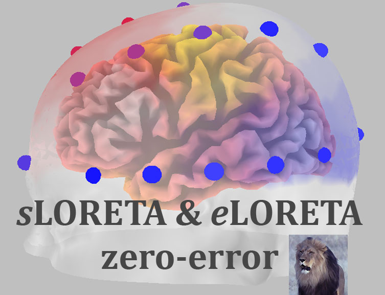
 We deal here with the EEG/MEG neuroimaging problem: given measurements of scalp electric potential differences (EEG: electroencephalogram) and extracranial magnetic fields (MEG: magnetoencephalogram), find the 3D distribution of the generating electric neuronal activity. This problem has no unique solution. Only particular solutions with “good” localization properties are of interest, since neuroimaging is concerned with the localization of brain function. A general family of linear imaging methods with exact, zero error localization to point-test sources can be found here. One particular member of this family is sLORETA (standardized low resolution brain electromagnetic tomography; Pascual-Marqui, Methods Find. Exp. Clin. Pharmacol. 2002, 24D:5-12; PDF file here). It is shown that sLORETA has no localization bias in the presence of measurement and biological noise. Another member of this family, denoted as eLORETA (exact low resolution brain electromagnetic tomography), is a genuine inverse solution (not merely a linear imaging method, nor a collection of one-at-a-time single best fitting dipoles) with exact, zero error localization in the presence of measurement and structured biological noise.
sLORETA/eLORETA software update 2008-November-04: Brain connectivity (oscillatory brain transactions) Tutorial and Viewer
This new update contains:
1. An alpha-version software for viewing brain connectivity results. The measures of brain connectivity used here are properly corrected for volume conduction and low resolution (http://arxiv.org/abs/0711.1455).
2. A tutorial on how to perform a brain connectivity analysis using eLORETA.
3. All tutorials (General, BRL-sLORETA norms, and Brain connectivity) are accessible from the Start menu.
4. A large number of minor bug fixes.
sLORETA/eLORETA software update 2008-August-23: Microstates, ICA, and BRL sLORETA norms 2008
This new update contains:
1. Lehmann's Brain Microstate Segmentation:
1.1. Lehmann, D. and Skrandies, W.: Reference-free identification of components of checkerboard-evoked multichannel potential fields. Electroenceph. Clin. Neurophysiol. 48: 609-621 (1980).
1.2. Lehmann, D., Ozaki, H. and Pal, I.: EEG alpha map series: brain micro-states by space-oriented adaptive segmentation. Electroenceph. Clin. Neurophysiol. 67: 271-288 (1987).
The Microstates are estimated as described in:
1.3. Pascual-Marqui, R.D., Michel, C.M. and Lehmann, D. Segmentation of brain electrical activity into microstates: model estimation and validation. IEEE T. Bio-Med. Eng. 42: 658-665 (1995).
2. Independent component analysis (ICA) / Blind source separation (BSS). Here you will find a family of algebraic ICA methods for EEG/ERP data (single subject and group data).
3. BRL – sLORETA norms 2008: The norms, the software, and their validation have been developed in collaboration with E. Roy John, Leslie Prichep, and Roberto Isenhart, from the Brain Research Laboratories (BRL), Department of Psychiatry, NY University School of Medicine, NY, USA.The main original paper on the methodology of the use of norms in EEG is: E.R. John, B.C. Karmel, W.C. Korning, P. Easton, D. Brown, H. Ahn, M. John, T. Harmony, L. Prichep, A. Toro, I. Gerson, F. Bartlet, R.W. Tatcher, H. Kaye, P. Valdés and E. Schwartz. Neurometrics: the use of numerical taxonomy to evaluate brain function. Science 196 (1977), pp. 1393–1410. These tools extend the NEUROMETRIC methodology to computed intracraneal sources using sLORETA.
4. The Microstate and ICA modules contain help files. The BRL sLORETA norms 2008 contains and extensive Tutorial.
sLORETA/eLORETA software update 2008-April-03
This new update contains:
1. Time-varying cross-spectral analysis and viewer. This can be applied to average ERPs or to average-removed single trial ERPs.
2. Format converter for binary time-varying cross-spectra to text.
3. ROI extractor. Regions of interest can now be extracted from sLORETA files, allowing users to make their own analyses on ROI data. Due to previous lack of documentation (now there is a bit more), some Users erroneously attempted to compute sLORETA for ROIs using the Utilities module "EEGs/ERPs to sLORETA" together with the special ROI transformation matrix (*.ROIspinv).
4. Computation of time-varying sLORETA spectra.
5. Brain connectivity measures (linear, nonlinear, total, instantaneous, and lagged) in the time-frequency domain.
6. Documentation has been added to many of the Utilities modules
7.
Documentation has been added to the statistical module (the Help button works).
8. The sLORETA viewer program includes in the slice-viewer an option to find and list all suprathreshold voxels.
Instantaneous and lagged measurements of linear and nonlinear dependence between groups of multivariate time series: frequency decomposition [ arXiv: 0711.1455 [stat.ME], 2007-November-09, http://arxiv.org/abs/0711.1455]
Measures of linear dependence (coherence) and nonlinear dependence (phase synchronization) between any number of multivariate time series are defined. The measures are expressed as the sum of lagged dependence and instantaneous dependence. The measures are non-negative, and take the value zero only when there is independence of the pertinent type. These measures are defined in the frequency domain and are applicable to stationary and non-stationary time series. These new results extend and refine significantly those presented in a previous technical report (Pascual-Marqui 2007, arXiv:0706.1776 [stat.ME], http://arxiv.org/abs/0706.1776 ), and have been largely motivated by the seminal paper on linear feedback by Geweke (1982 JASA 77:304-313). One important field of application is neurophysiology, where the time series consist of electric neuronal activity at several brain locations. Coherence and phase synchronization are interpreted as “connectivity” between locations. However, any measure of dependence is highly contaminated with an instantaneous, non-physiological contribution due to volume conduction and low spatial resolution. The new techniques remove this confounding factor considerably. Moreover, the measures of dependence can be applied to any number of brain areas jointly, i.e. distributed cortical networks, whose activity can be estimated with eLORETA (Pascual-Marqui 2007, arXiv:0710.3341 [math-ph], http://arxiv.org/abs/0710.3341 ).
sLORETA/eLORETA software update 2007-November-15
This version (2007-November-15) adds:
(1) The new eLORETA method
(2) Tools for defining cortical regions of interest (ROIs)
(3)
New instantaneous and lagged measurements of linear and nonlinear dependence between groups of multivariate time series. Connectivity is computed between cortical regions of interest (ROIs).
(4) Statistical tools (SnPM methodology) for hypothesis testing on the new connectivity measures
eLORETA paper (2007-Oct-23)
A technical report with new results in EEG/MEG-neuroimaging (including eLORETA) can be downloaded from http://arxiv.org/abs/0710.3341
Cite as: "R.D. Pascual-Marqui: Discrete, 3D distributed, linear imaging methods of electric neuronal activity. Part 1: exact, zero error localization. arXiv:0710.3341 [math-ph], 2007-October-17, http://arxiv.org/pdf/0710.3341"
Abstract: This paper deals with the EEG/MEG neuroimaging problem: given measurements of scalp electric potential differences (EEG: electroencephalogram) and extracranial magnetic fields (MEG: magnetoencephalogram), find the 3D distribution of the generating electric neuronal activity. This problem has no unique solution. Only particular solutions with “good” localization properties are of interest, since neuroimaging is concerned with the localization of brain function. In this paper, a general family of linear imaging methods with exact, zero error localization to point-test sources is presented. One particular member of this family is sLORETA. It is shown here that sLORETA has no localization bias in the presence of measurement and biological noise. Another member of this family, denoted as eLORETA (exact low resolution brain electromagnetic tomography), is a genuine inverse solution (not merely a linear imaging method) with exact, zero error localization in the presence of measurement and structured biological noise. The general family of imaging methods is further extended to include data-dependent (adaptive) quasi-linear imaging methods, also with the exact, zero error localization property.
sLORETA software update 2007-March-20
This version (2007-March-20) adds four new items to the sLORETA utilities module:
(1) Old LORET-KEY electrode coordinates can be registered to the MNI152 scalp. This is achieved by a spline that projects the electrodes onto the scalp with minimum distortion.
(2) Realistic electrode coordinates can now be registered to the MNI152 scalp. Coordinates for the following landmarks are required: nasion, inion, right and left preauriculars (anterior root of the center of the peak region of the tragus), and Cz (half-way Nz to Iz). All units in millimeters. In a first step, a 12-parameter affine transformation is used, followed by a spline that projects the electrodes onto the scalp with minimum distortion.
(3) Electrodes can be viewed in three orthogonal slices in the MNI152 template. This may be used for visual inspection of registered electrodes.
(4) In previous versions, the transformation matrix was calculated using the lead field for fixed electrode positions only, which was a crude approximation for realistic electrodes. In this version much better results are achieved with a spline interpolation for any electrode coordinates. Advise: don't use non-cephalic recording electrodes such as cheeks, eyes, neck, etc.
LORETA-KEY group created for the announcement of new software, updates, upgrades (2007-Feb-09)
Please join the group. While subscribing, make sure you click the link “Edit my membership”, and under the heading “How do you want to read this group?”, select one of the email options (otherwise, you should check periodically the group discussion on the web).