Navigation auf uzh.ch
Navigation auf uzh.ch
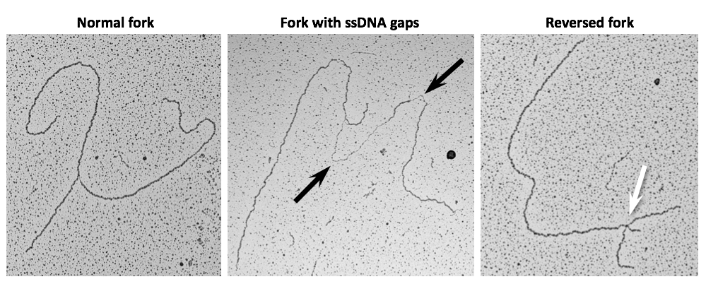
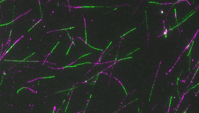
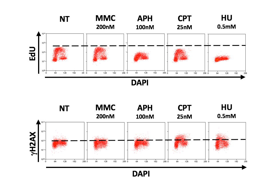
Interference with DNA replication is a prominent clinical strategy to counteract uncontrolled cancer cell-proliferation, with the rationale that the majority of non-cancerous cells is quiescent and thus remains largely unaffected. A major limitation for effective cancer therapy however is the limited knowledge on the mechanism of action of most chemotherapeutic regimens. Understanding how the replication apparatus is specifically challenged by each treatment and which factors assist the cellular response to these perturbations is instrumental to any strategy aiming to improve cancer chemotherapy and to limit chemoresistance, an outcome frequently observed in cancer patients. To that aim, we successfully refined specialized single-molecule approaches on replication intermediates and pioneered their use on cancer cell lines to uncover molecular responses to drug-induced replication stress. Thereby we have provided the first direct visualization of replication fork remodelling into four-way junctions (namely fork reversal) in human cells, as a global, transient, evolutionarily conserved response to various perturbations of the replication process. These studies allowed refining long-standing models on the mechanism of action of commonly used anticancer drugs.
Main goal of this research line is the visualization and molecular characterization of cancer cells’ reaction to commonly used cancer chemotherapeutic treatments, therewith providing new targets and strategies to improve cancer therapy and address chemoresistance.
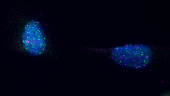
While continuing to exploit our powerful single-molecule approaches, we are currently expanding our portfolio of imaging approaches and molecular analyses to investigate at high resolution how the replication stress response is orchestrated spatiotemporally within the nucleus. We aim to decipher how essential components of nuclear architecture and dynamics – e.g. nuclear cytoskeleton, chromatin organization, 3D genome interactions - assist the replication apparatus facing replication stress. To do so, we are actively collaborating with top experts in visualization and functional analysis of nuclear/genome organization, probing the role of specific nuclear factors and signalling pathways in the replication stress and chemotherapy response.
A. Ray Chaudhuri, Y. Hashimoto, R. Herrador, K.J. Neelsen, D. Fachinetti, R. Bermejo, A. Cocito, V. Costanzo and M. Lopes (2012). Topoisomerase I poisoning results in PARP-mediated replication fork reversal. Nature Structural and Molecular Biology 19: 417–423.
R. Zellweger, D. Dalcher, K. Mutreja, J. A. Schmid, R. Herrador, M. Berti, A. Vindigni and M. Lopes (2015). Rad51-mediated replication fork reversal is a global response to genotoxic treatments in human cells. Journal of Cell Biology 208:563-79.
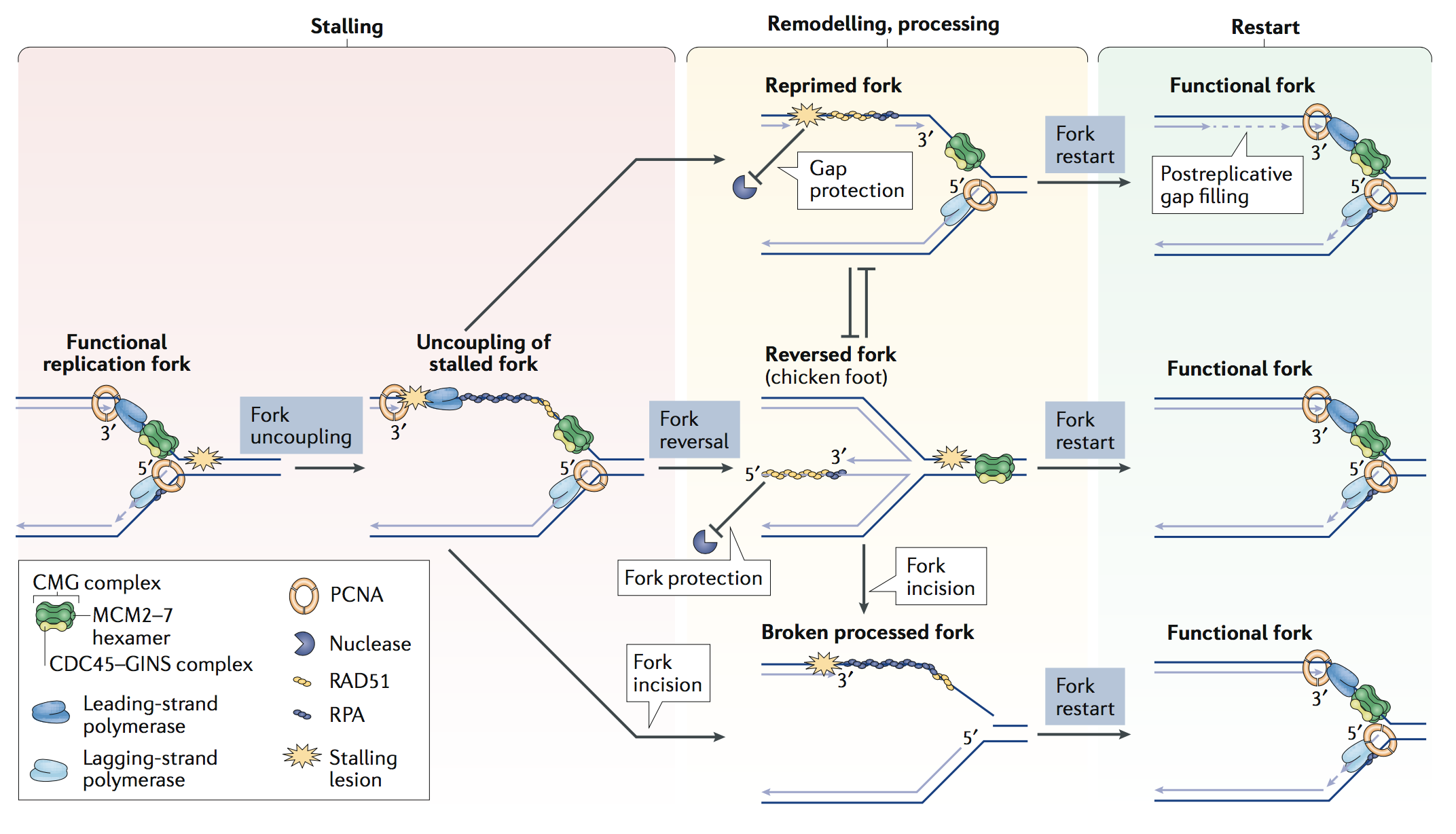
K. Neelsen and M. Lopes (2015). Replication fork reversal in eukaryotes: from dead end to dynamic response. Nature Reviews Molecular Cellular Biology, 16:207-220.
K. Mutreja, J. Krietsch, J. Hess, S. Ursich, M. Berti, F.K. Roessler, R. Zellweger, M. Patra, G. Gasser and M. Lopes (2018). ATR-Mediated Global Fork Slowing and Reversal Assist Fork Traverse and Prevent Chromosomal Breakage at DNA Interstrand Cross-Links. Cell Reports, 24(10):2629-2642.e5.
M. Berti, D. Cortez and M. Lopes (2020). The plasticity of DNA replication forks in response to clinically-relevant genotoxic stress. Nature Reviews Mol Cell Biol.