Navigation auf uzh.ch

Suche
  • 01_Schaf-OP

    The sheep that are used for this type of research are kept in small groups to give them social contacts and are cared for by a team of veterinarians. (Image: Frank Brüderli, UZH)

  • 02_Schaf-OP

    First, the wool is removed from the sheep’s neck and its skin is disinfected. (Image: Frank Brüderli, UZH)

  • 03_Schaf-OP

    The vet attaches a cannula to prepare for the infusion. This way, drugs can be administered without causing pain or stress to the animal. (Image: Frank Brüderli, UZH)

  • 04_Schaf-OP

    After the sheep has been sedated in the animal pen, it is transported to the operating room. (Image: Frank Brüderli, UZH)

  • 05_Schaf-OP

    A tube is placed in the trachea to maintain controlled anesthesia and ventilate the animal. To do this, the veterinarian first wash the sheep’s mouth. Pain treatment in sheep is comparable to that in humans. (Image: Frank Brüderli, UZH)

  • 06_Schaf_OP

    The sheep, which is now under anesthesia, is transferred to the operating table by two vets. (Image: Frank Brüderli, UZH)

  • 07_Schaf-OP

    Surgery is performed with the animal lying on its back and strapped to the table. (Image: Frank Brüderli, UZH)

  • 08_Schaf-OP

    Before surgery begins, the veterinary team examines the sheep’s uterus using ultrasound. (Image: Frank Brüderli, UZH)

  • 09_Schaf-OP

    Surgery is carried out under strictly sterile conditions to avoid infections – similar to surgery in humans. (Image: Frank Brüderli, UZH)

  • 10_Schaf-OP

    First, the surgeon opens up the sheep’s abdomen to expose the uterus. (Image: Frank Brüderli, UZH)

  • 11_Schaf-OP

    The pregnant uterus is exposed so that the next steps can be performed as precisely as possible. (Image: Frank Brüderli, UZH)

  • 12_Schaf-OP

    Depending on the scientific research question, treatment of the membranes surrounding the developing embryo is performed in open surgery or with minimal invasion. (Image: Frank Brüderli, UZH)

  • 13_Schaf-OP

    Surgical procedures on sheep are performed by experienced surgeons using state-of-the-art equipment. (Image: Frank Brüderli, UZH)

  • 14_Schaf-OP

    The ruptured membrane is sealed tight using a “plaster”. The tools required for this were developed by UZH spin-off KOVE medical AG. (Image: Martin Ehrbar, UZH)

  • 15_Schaf-OP

    The procedure is monitored using a fetoscope to systematically adapt and refine it. Such cameras are also used for minimally invasive procedures inside the human amniotic sac. (Image: Martin Ehrbar, UZH)

  • 16_Schaf-OP

    After the surgical procedure, the “plaster” is applied through the same cannula through which surgery was performed. Fetoscope images show a folded umbrella-like container being pushed into the amniotic sac. (Image: Martin Ehrbar, UZH)

  • 17_Schaf-OP

    The umbrella-like container is applied onto the rupture and attached to the membrane using a tissue adhesive. Novel adhesives are currently being developed that specifically stimulate the healing process of the membranes. (Image: Martin Ehrbar, UZH)

  • 18_Schaf-OP

    After the operation, as the sheep gradually wakes up from the anesthesia, it is transported back to the pen by two vets. (Image: Frank Brüderli, UZH)

  • 19_Schaf-OP

    Back in the animal pen, the sheep first gets some food – under close supervision by the veterinarian. (Image: Frank Brüderli, UZH)

  • 20_Schaf-OP

    The animal caretakers and vets continuously monitor the health and wellbeing of the sheep, and record any steps and measures taken. (Image: Frank Brüderli, UZH)

Preventing Premature Births after Fetal Surgery

Operations on unborn babies with severe conditions can save children’s lives and enable them to lead better lives. Unfortunately, this kind of surgery may induce the rupture of the fetal membranes, which in many cases leads to preterm births. Bioengineer Martin Ehrbar and physician Nicole Ochsenbein-Kölble are developing new methods to repair defective membranes – not least thanks to research involving sheep.

Babies born with spina bifida may suffer from limited mobility, incontinence and hydrocephalus. In rare cases, the fetuses of identical twins sharing a placenta may share unequal amounts of the placenta’s blood supply. Both of these conditions may lead to lifelong physical and mental impairments for the affected children, or even death of the babies in the womb. The survival chances and quality of life of these children can be significantly improved through fetal surgery. These advances have been made possible in no small part thanks to research involving animals, especially sheep.

Reducing preterm births by repairing membrane defects

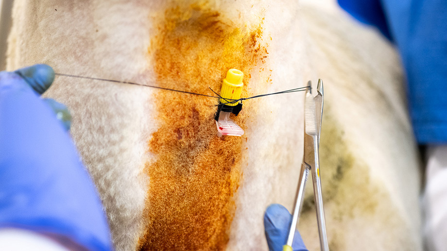
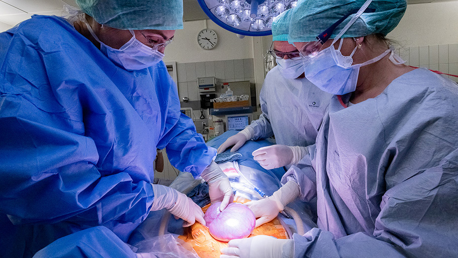
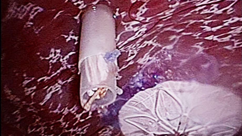
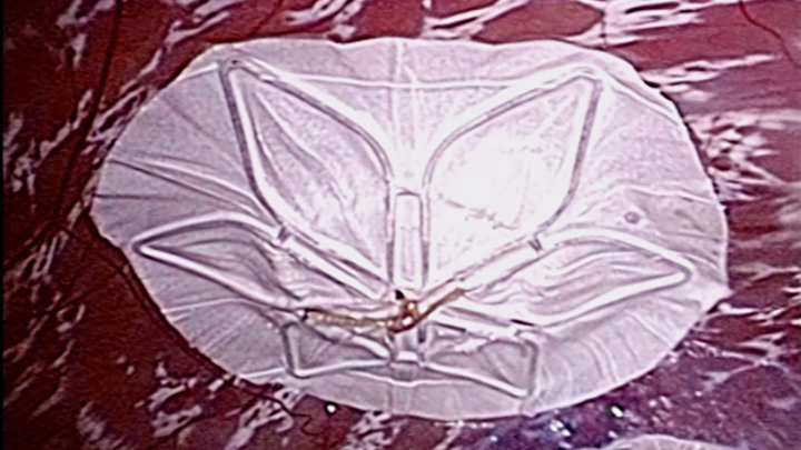
In many cases, prenatal surgery causes the babies to be born early, since this kind of procedure may induce the rupture of the membranes surrounding the fetus. This can cause serious complications for the newborns. Martin Ehrbar, professor of fetal healing and tissue engineering at UZH, and Nicole Ochsenbein-Kölble, Director at the Department of Obstetrics at the University Hospital Zurich, have been investigating a solution to prevent preterm births for years. They are developing biomaterial-based treatments that immediately seal ruptured membranes and promote wound healing. This “plaster” looks like an umbrella-like container that can be opened and closed.

Bespoke biomaterials for regenerative medicine

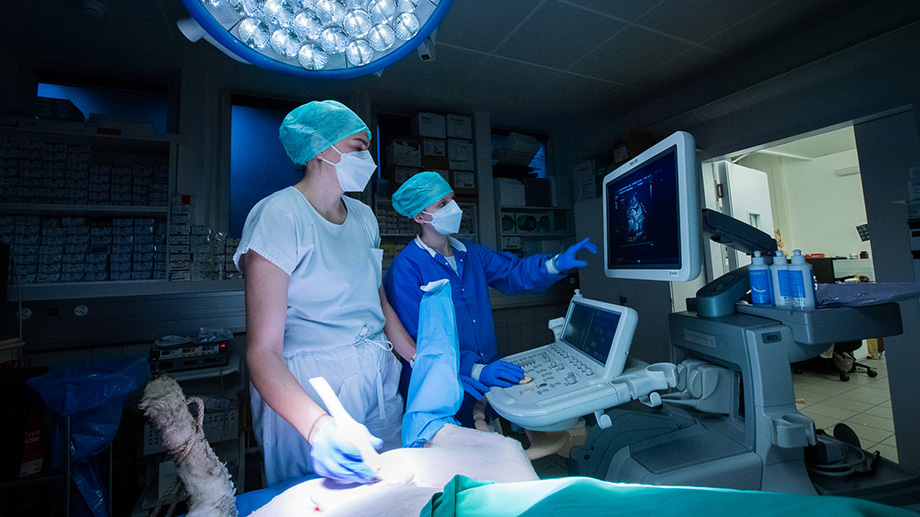
The novel synthetic biomaterials developed by Ehrbar and Ochsenbein-Kölble stabilize the membrane to facilitate cell growth. This makes it possible to produce tissues that mimic those of human fetal membranes. The researchers are investigating the biomaterials’ ability to support the healing of defective membranes by using cell cultures as well as pregnant sheep. Using tissue cultures as a model of the pregnant uterus during surgery, the UZH scientists have developed tools to apply the healing biomaterials to the ruptured membrane as gently as possible.

Biological complexity calls for animal testing

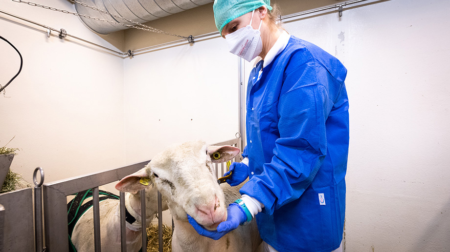
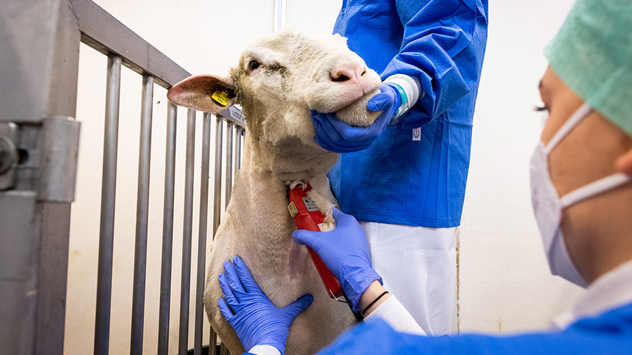
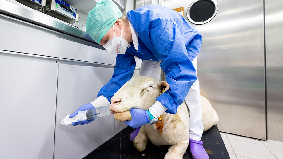
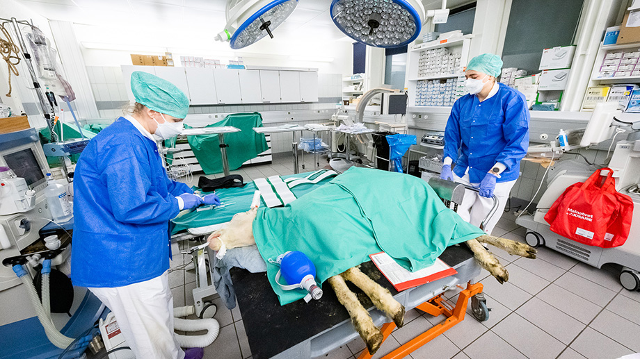
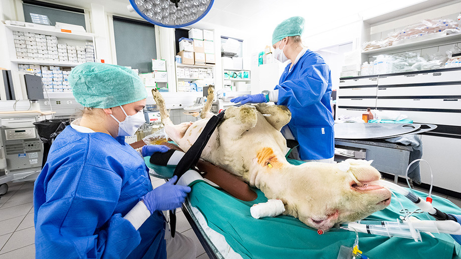
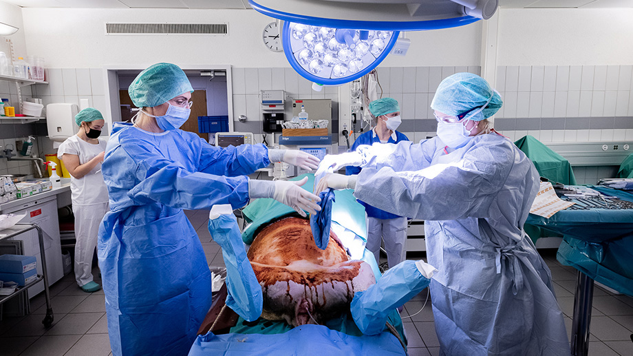
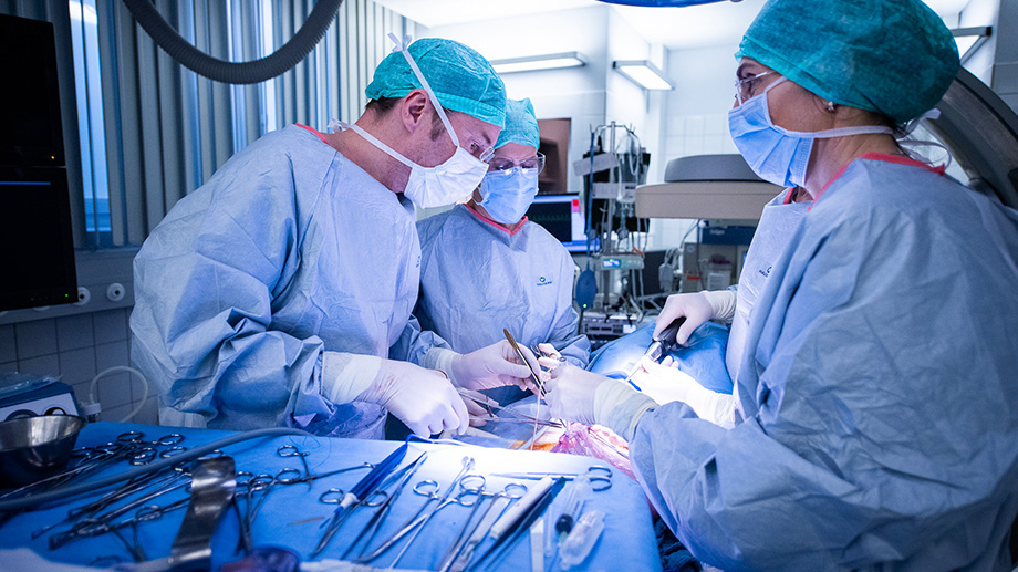
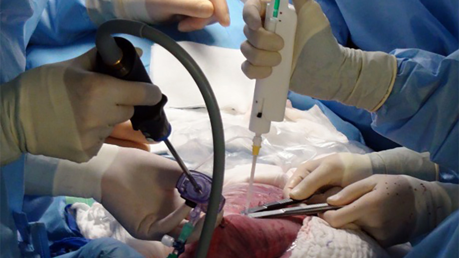
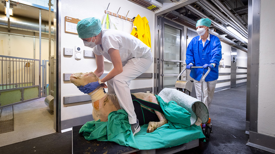
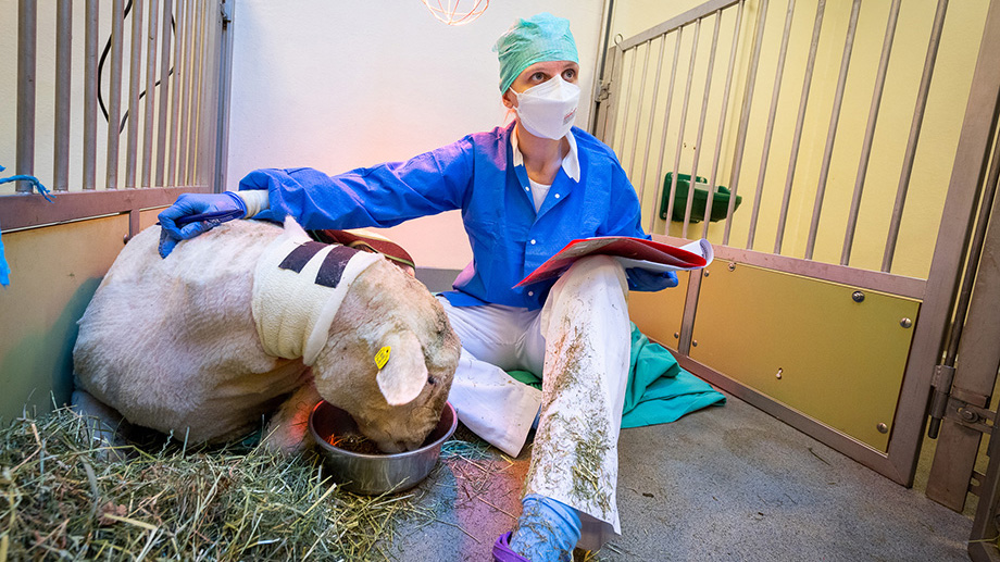
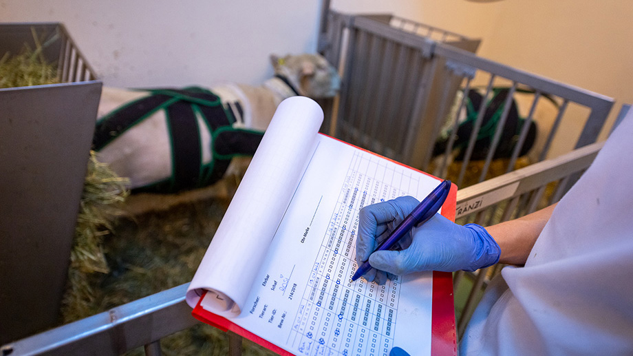
One of the advantages of using tissue cultures is that they enable researchers to conduct in-depth studies, while significantly reducing the number of animal experiments. However, the effects of amniotic fluid on the biomaterials and the healing processes are so complex that the only way to examine the treatment’s effectiveness in humans is to use live animals. Ehrbar and Ochsenbein-Kölble have thus developed an animal model in pregnant sheep to test fetal membrane healing through biomaterials.

Repairing membrane defects in animal models

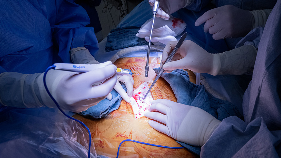
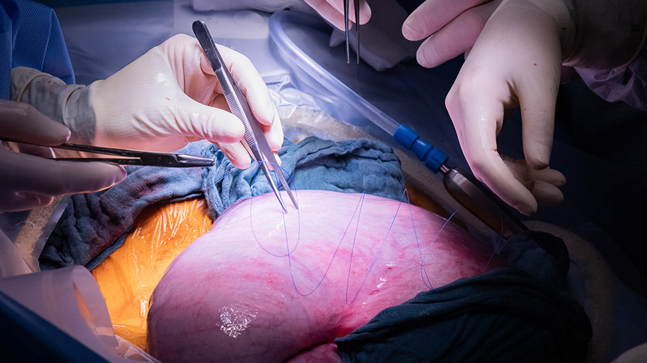
Comparable to the procedures on human fetuses, minimal invasive surgery is performed on pregnant ewes under anesthetic to open the uterus and fetal membranes. Martin Ehrbar and Nicole Ochsenbein-Kölble are supported by veterinarian Miriam Weisskopf from the Center for Surgical Research at UZH. The resulting membrane defects are then sealed using the newly developed biomaterials. Tissue analyses have confirmed that initial results on healing membrane defects in sheep are promising. The biomaterials are now being further optimized for future use in humans.

Weiterführende Informationen

Ehrbar research group

Prof. Dr. sc. nat. Martin Ehrbar
Research Group "Biomaterials and Regenerative Medicine"
Department of Obstetrics, UZH/USZ

Ochsenbein-Kölble research group

Prof. Dr. med. Nicole Ochsenbein-Kölble
Research Group "Multiples, fetoscopic laser therapy and fetal surgery" and "Biology of fetal membranes"
Department of Obstetrics, UZH/USZ

Center for Surgical Research

Dr. med. vet. Miriam Weisskopf
Anaesthesia and Peri-Operative Pain Research
Center for Surgical Research, UZH/USZ