Navigation auf uzh.ch
Navigation auf uzh.ch
We study the properties of disordered and heterogeneous systems out of equilibrium. This encompasses light transport in turbid media and photonic glasses, with applications in imaging, structural colours, light harvesting for energy and secure optical communication. A second focus of our activities is the study of the elastic properties of growing biological tissues and their influence on development and pattern formation, e.g. in embryonal development of drosophila, such as ventral furrow formation or dorsal closure. In all these fields our investigations are mainly experimental, developing the tools necessary, e.g. to study forces in tissues on the scale from nN to mN, however we also use computational modeling to guide these experiments.
Modelling Ventral Furrow Formation in Drosophila embryos
The invagination of mesoderm during ventral furrow formation in the Drosophila embryo is part of the archetypal morphogenic process of gastrulation. This is one of the most fundamental processes during embryonal development, where the initial symmetry breaking of the embryonal shell takes place. To explore the roles of the active cellular forces and the regulation of these forces, we developed an integrated vertex model that gives a complete description of the process.
In this, it combines the regulation of morphogen expression in a regulatory network with cell movements and the corresponding tissue mechanics. Finally, in order to take into account mechanical regulation of biological development, the regulatory network is influenced by the mechanical tensions, thus providing a mechanism for mechanical feedback. In our simulations, a successful furrow formation requires an apical tension gradient, decreased basal tension, and increased lateral tension, which corresponds to apical constriction, basal expansion, and apicobasal shortening respectively. These tensions can be dynamically obtained from a regulation by the morphogen twist, which acts as a regulator of myosin, responsible of contractile tension in the cell boundaries.
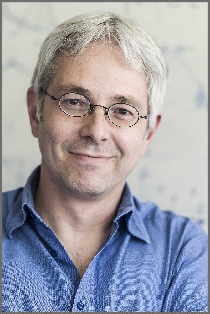
Figure: A) A simulation of normal invagination using the regulatory network to control the expression of twist determining the tensions in the cells. These tensions are colour coded using the scale shown on the right.
B) A simulation using the same regulatory network, however here the embryo has been constantly deformed using an external compression. Such a deformation experimentally leads to the overexpression of twist and thus constricts invagination. This is also seen in the simulations shown here.
Our regulatory equations can reproduce this dynamics of the the apical tension gradient, basal tension decrease, and lateral tension increase, which are corresponding to apical constriction, basal expansion, and apico-basal shortening respectively. Then we integrated the regulation network and the vertex model. The results show that our integrated model is also able to form a closed ventral furrow although there are anomalous deformations in the intermediate states. Our model also replicates the ectopic twist expression induced by mechanical compression. The ectopic expression of twist prevents mesoderm invagination, which is consistent with experimental observations (see figure). Our model also predicts the ectopic invagination in presence of a locally applied external force gradient, which has not been verified in vivo.
Highlighted Publications:
1. An integrated vertex model of the mesoderm invagination during the embryonic development of
Drosophila,
J. Jiang and C.M. Aegerter, Journal of theoretical Biology 572, 111581 (2023).
We are conducting research and development in Medical Physics, Theoretical Biology and Medical Modelling. Our main topics are: Development of radio-biological models, space radiation research, Monte Carlo simulations and dosimetry for radiotherapy and imaging and the development of novel detector systems.
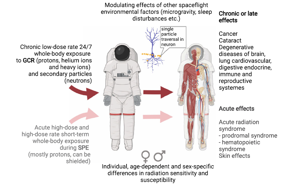
In 2023 our work was focused on Space Radiation Research (Figure). One project, in collaboration with CERN, is aiming on the development of a beam line to simulate high-LET irradiation of the cosmic galactic background for earth based material and biological experiments. In a second project, we are developing an European risk assessment software to assess organ dose equivalents for European astronauts. For Astronauts the probability of surviving cancer free a Mars swing by mission until retirement age is being reduced by a range from 0.78% to 2.63%. It was found that the risks for females are higher than for males, and the risks at solar minimum are higher than at solar maximum.
Figure: Radiation exposure during space missions beyond low Earth orbit and health effects of space radiation (from [1]).
Highlighted Publications:
Radiotherapy is one of the mainstays of cancer treatment and a highly technology-driven field of medicine. Our research group contributes to the development of radiotherapy technology by applying concepts from physics, mathematics, statistics, and machine learning to problems in medical imaging and radiation oncology.
We focus on three areas of research:
1) Radiotherapy treatment planning: We work on mathematical optimization methods to optimally combine x-ray and proton beams, and to optimally distribute radiation dose over multiple treatment days and radiation modalities [1].
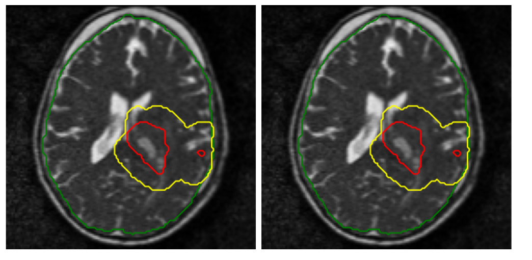
2) Target delineation and outcome prediction: Here, we focus on quantitative modeling of tumor progression and the analysis of medical images such as MRI, CT, and PET, with the goal of precisely defining the region to be irradiated and predicting the patient’s response to treatment (see Figure).
3) Latest radiotherapy technology: We perform research on FLASH therapy and MR-guided therapy at the MR-Linac, a combination of MRI scanner and radiotherapy device allows MR imaging of a patient during treatment (see Figure).
Figure: MR-images of a brain tumor patient, showing tumor changes between the start (left) and the end (right) of treatment.
Highlighted Publications:
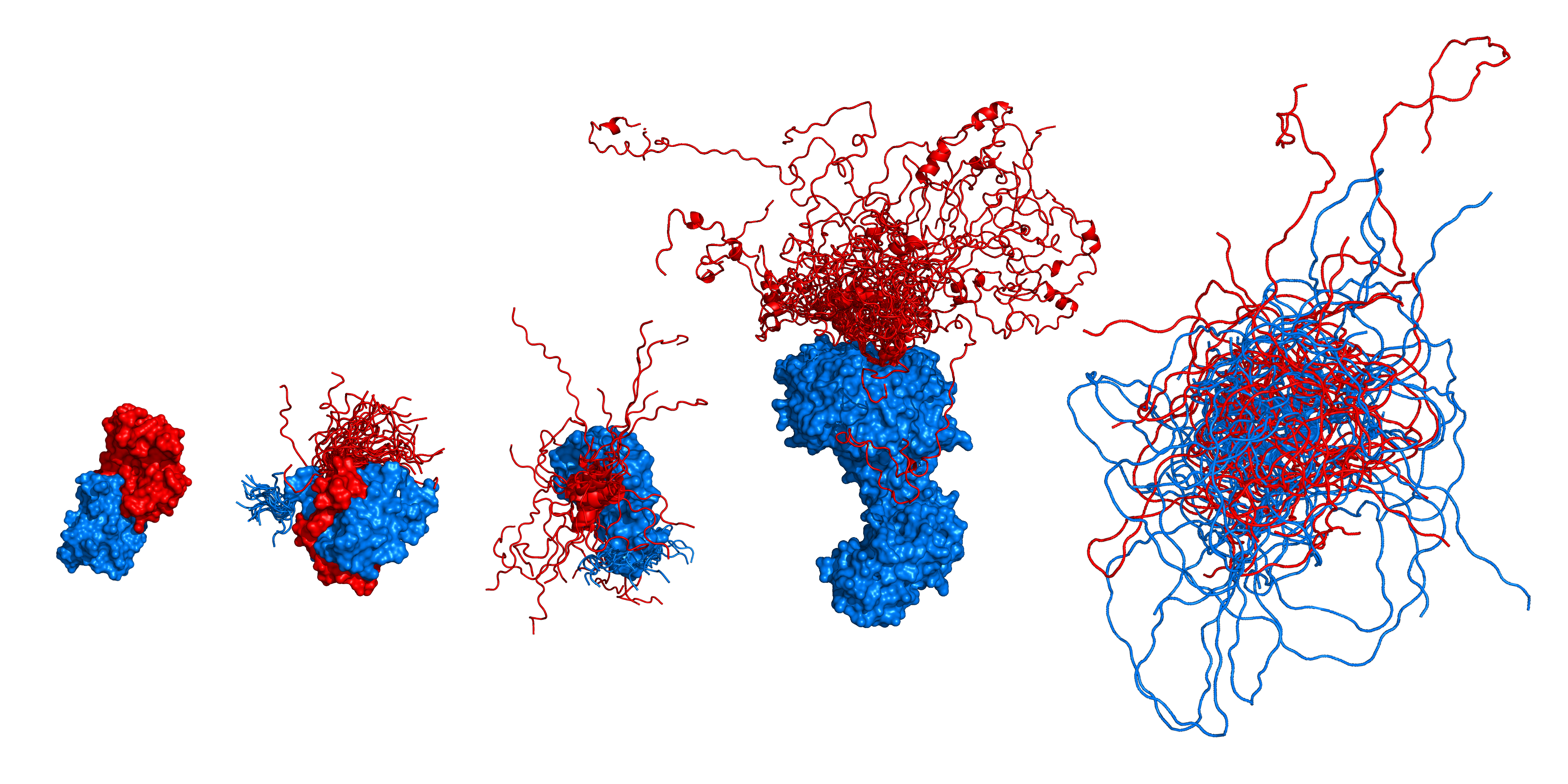
We study the structure, dynamics, and functions of biomolecules, especially proteins, the nanomachines of life. Towards this goal, we develop and apply single- molecule fluorescence and force spectroscopy, often in close combination with theory and simulations. A particularly important tool is Förster resonance energy transfer (FRET), a spectroscopic nanoscale ruler.
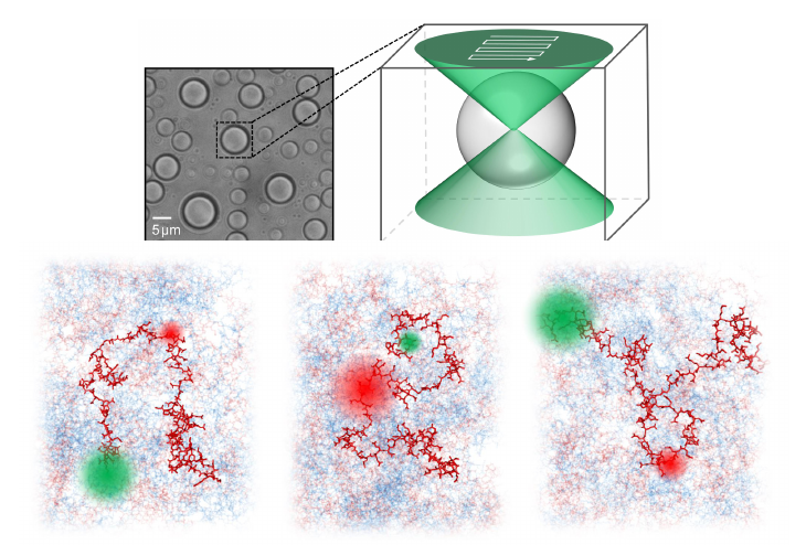
Two highlights were the development of a droplet-based microfluidic mixing device for the rapid triggering of biomolecular reactions [1], and the single-molecule study of nanoscopic dynamics within phase-separated droplets of biomolecular polyelectrolytes and their interpretation based on massive molecular dynamics simulations [2].
Figure: Confocal single-molecule FRET measurements in complex coacervates of highly charged protein molecules revealed polymer dynamics on sub-microsecond timescales. The snapshots at the bottom illustrate the behavior of fluorescently labeled molecules in a dense phase of protein.
Highlighted Publications:


